
Some anatomy for the basic forearm radiographs! Kevin GrepMed
The trauma dated of 1 day and X-ray was initially judged normal in the emergency department. Due to the persistence of the pain and the functional impotence, the patient presented again to our.

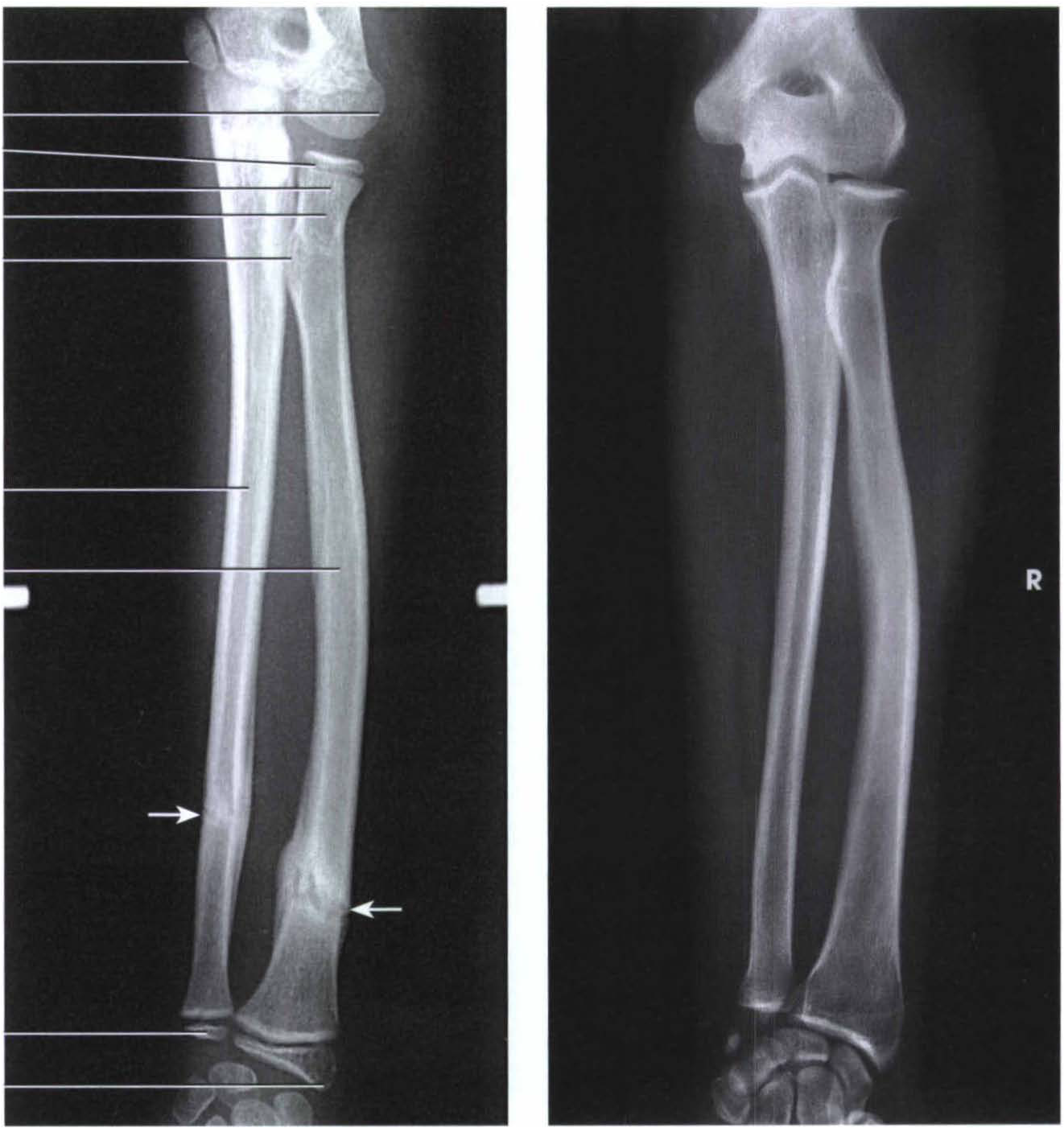
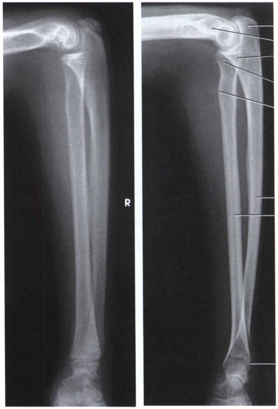
Forearm X Ray Anatomy
Hand, humeral and antebrachii X-ray showed no fracture nor dislocation. Electromyography (EMG) test showed no nerve conduc- tion velocity of left axillary nerve injury. Patient underwent physio- therapy and given neuroprotectors. After 7 days, there was a significant improvement in shoulder abduction and left arm prona- tion.
Gudang Medis teknik radiografi antebrachii
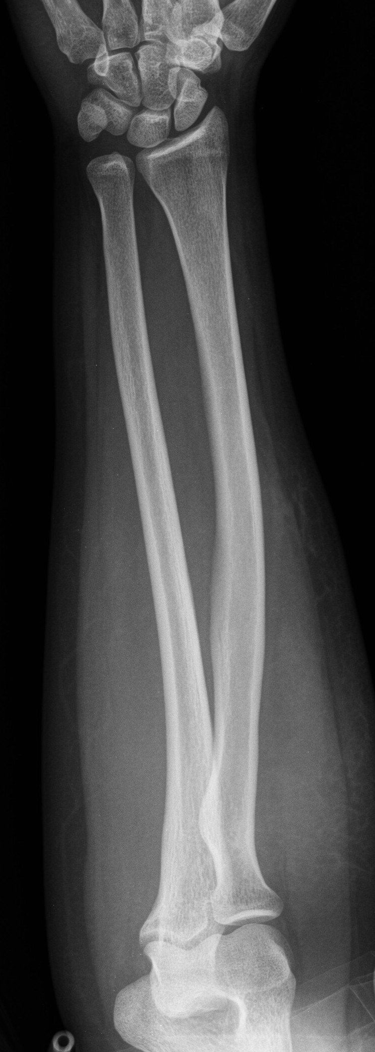
Presentation Fall from bike. Pain in wrist. Patient Data Age: 9 years Gender: Male x-ray Frontal Lateral Normal examination. No fracture. No joint effusion. Case Discussion Forearm x-rays are difficult. You end up with an frontal view of the wrist and a lateral view of the elbow on one image and the opposite on the other.
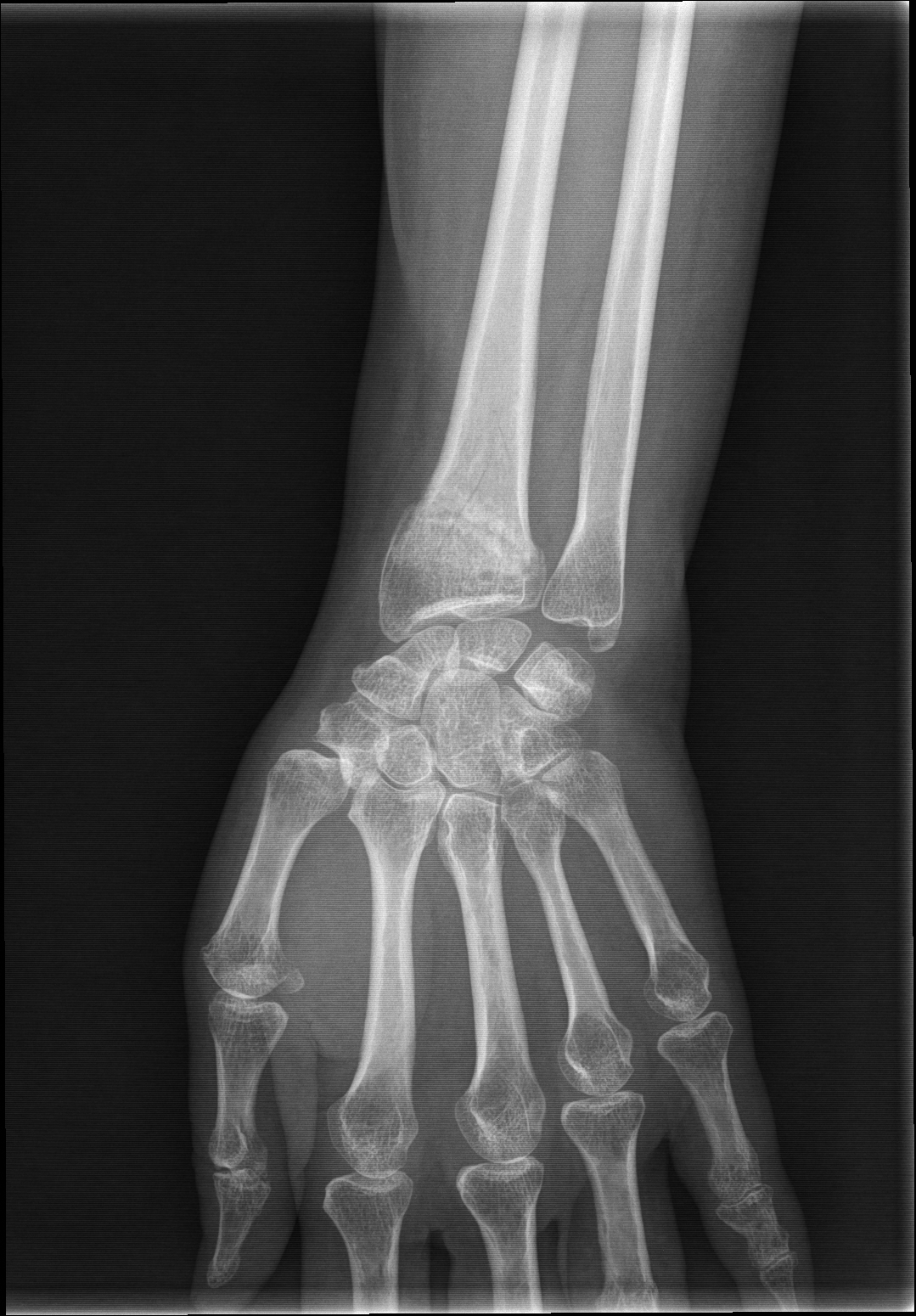
X Ray Wrist Joint Post Trauma Radiology Imaging
Imaging of the body is often complicated by the fact that anatomic structures overlap each other. Diagnostic accuracy of radiographs generally refers to how well an exam can predict the presence (or absence) of a disease or condition. The technologist plays a pivotal role in improving diagnostic accuracy by providing diagnostic images.[1] This requires a technologist to be aware of the various.
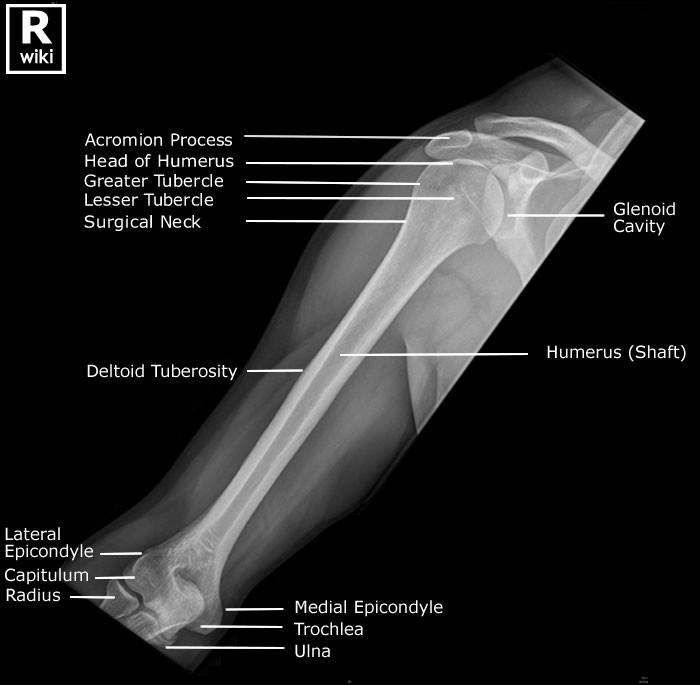
Humerus Radiographic Anatomy wikiRadiography
This view demonstrates the elbow joint in its natural anatomical position allowing for assessment of suspected dislocations or fractures and localizing foreign bodies within the forearm. patient is seated alongside the table forearm is supinated, and its dorsal surface is kept in contact with the cassette with extension at the elbow joint
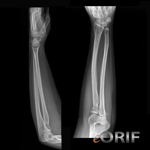
Forearm Xray eORIF
Small Animal Elbow and Antebrachium Radiography Issue: July/August 2012 This is the sixth article in our Imaging Essentials series, which is focused on providing comprehensive information on radiography of different anatomic areas of dogs and cats. The following articles are available at todaysveterinarypractice.com:

Radiographs of the left elbow and forearm at 6month followup
Radiographic features Forearm fractures are readily diagnosed on plain radiographs, and further imaging is rarely required. Plain radiograph AP and lateral X-rays of the forearm are performed. A radial or ulnar fracture will be visible on at least one view.

AB AP and lateral radiographs showing the original proximal radius
Radius and ulnar shaft fractures, also known as adult both bone forearm fractures, are common fractures of the forearm caused by either direct trauma or indirect trauma (fall). Diagnosis is made by physical exam and plain orthogonal radiographs. Treatment is generally surgical open reduction and internal fixation with compression plating of.

Image
Anatomy . The brachialis muscle originates from the front of your humerus, or upper arm bone.It arises from the distal part of the bone, below your biceps brachii muscle. It then courses down the front of your arm, over your elbow joint, and inserts on the coronoid process and tuberosity of your ulna.The brachialis muscle, along with the supinator muscle, makes up the floor of the cubital.

Forearm Radiograph Radiology student, Medical radiography, Medical
X-rays are taken to ensure that the reduction was successful. The cast is usually maintained for about 6 weeks. X-ray in cast. Unsuccesful reduction. Guidelines for non-acceptable reduction are (8): Radial shortening > 5 mm; Radial inclination Tilt on lateral projection > 10 degrees dorsal tilt and > 20 degrees volar tilt;
AP (A) and lateral (B) forearm xray of patient 3, showing a dislocated
Also called ambient cistern is a cistern of the subarachnoid space between the posterior end of the corpus callosum and the superior surface of the cerebellum. It is sometimes defined as including the quadrigerminal cistern. On the left a coronal view of the segments of the middle cerebral artery. Horizontal M1-segment.

Pin by Tracey Burns on Radiology Diagnostic imaging, Radiology
Both Bone Forearm Fractures are one of the most common pediatric fractures, estimated around 40% of all pediatric fractures. Diagnosis is made with plain radiographs of the forearm. Treatment is closed reduction and casting for the majority of fractures. Surgical intervention is indicated for significantly displaced or angulated fractures in.

Interpreting Elbow and Forearm Radiographs — Taming the SRU
Publicationdate 2005-08-23. This article is based on a presentation given by Louis Gilula and adapted for the Radiology Assistant by Ileana Chesaru. First a systematic analysis of the wrist is presented to look for carpal instability and fracture dislocation. Secondly cases are presented as examples in the chapter systematic review and diagnosis.
Gudang Medis teknik radiografi antebrachii
OBJECTIVE. The purpose of this article is to review the anatomy, biomechanics, and multimodality imaging findings of common and uncommon distal radioulnar joint (DRUJ), triangular fibrocartilage complex, and distal ulna abnormalities. CONCLUSION. The DRUJ is a common site for acute and chronic injuries and is frequently imaged to evaluate chronic wrist pain, forearm dysfunction, and traumatic.
UCSD Musculoskeletal Radiology
Forearm x-rays are indicated for a variety of settings including: trauma bony tenderness suspected fracture obvious deformity non-traumatic pain suspected foreign body Projections Standard projections anteroposterior view demonstrates the radius and the ulna in the natural anatomical position lateral view projection 90° to the AP view

Show Me A Picture Of A XRay imgultra
fracture location (extra-, juxta- or intra-articular) degree of angulation degree of displacement carpus ensure no carpal malalignment or fractures are present assess articulation of radio-lunate and radio-scaphoid joint
